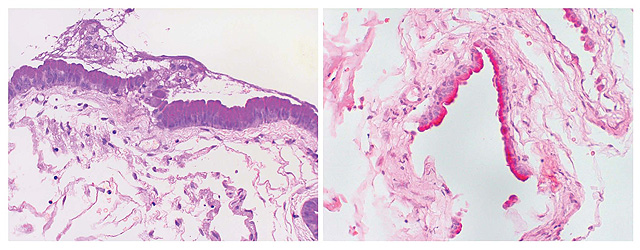
Figure 4 (Left) Periodic Acid Shift (PAS) Stain (right) Mucin Stain
PATHOLOGY
The pathology findings indicate the real origin of these lesions, showing an
epithelium of digestive or respiratory type lining the cavity of the cyst. A
fibrous capsule, variable in thickness, is always present and the cysts are
lined by epithelium varying from cuboidal to columnar and occasionally
pseudo-stratified type . The epithelium is strongly PAS positive and rests on a
connective tissue base. A varying number of globet cells are present within the
epithelium and are responsible for the mucinous content. (8).Electron microscopy
can reveal areas of ciliated epithelium whereas non ciliated cells wich
microvilli are present in every case with characteristic surface granular
glycocalyx coating. They possess features of secretory function, having
prominent nuclei, and, as well, tight junctions are present (9).
In our patient, a
typical fragment of foregut epithelium was found with secretory products: the
secretory function gives support to the possibility of secretion of contrast
inside the lesion, a fact never seen and described before in literature. In
addition several calcifications have been found.
REFERENCES
-
Agnoli AL, Laun A, Schonmyr R.
Enterogenous intraspinal cyst. J Neurosurg 1984;61:824-840.
-
Rodaci MA, Teixeira WR, Boer
VHT, et al. Intradural extramedullary high cervical neurenteric cyst.
Neuroadiology 1987; 29:588.
-
Shakudo M, Inoue Y,Ohata K,
Tanaka S. Neurenteric Cyst with Alteration of Signal Intensity on Follow-up MR
Images. AJNR Am J Neuroradiol 2001: 22;496-498.
-
Naidich TP, Mclone DG. Growth
and development. In Kricum ME. Imging Modalities in spinal disordes, pp.1-19.
Philadelphia, WB Saunders, 1988.
-
Brooks BS, Duvall ER, El
Gammal T, et al. Neuroimaging features of neurenteric cyst: analysis of nine
cases and rewiew of literature. AJNR Am J Neuroradiol 2001 May
1993;14:735-746.
-
Martin AJ, Penney CC. Spinal
neurenteric cyst. Arch Neurol 2001; 58:126-127.
-
Pierot L, Dormont D, Oueslati
S, et al. Gadolinium-DTPA enhanced Mr imaging of intradural neurenteric cyst.
J Comput tomogr 1988; 12(5):762-764.
-
Russell D, Rubinstein LJ.
Pathology of tumors of nervous system. 5th edn. Baltimore: Williams et
Wilkins; pp704-705, 690-695.
-
Elmadbouth H, Halpin SFS,
NealJ et al. Posterior fossa ephithelial cyst: case report and review of the
literature. AJNR Am J Neuroraidiol 2001 20:681-685.
-
Kallmes DF, Provenzale JM,
Cloft H et al. Typical and atypical MR imaging features of intracranial
eoidemoid tumor. AJR Am J Roentgenol 169:883-887.
-
Ochi M, Hayashi K, Hayashi T,
et al. Unusual CT and MR appearance of an epidermoid tumor of the
cerebellopontine angle. AJNR Am J Neuroradiol 1998;19:1113-1115.
-
Tsuruda JS, Chew WM, Moseley
ME, Norman D. Diffusion-weighted MR imaging of the brain: value of
differentiating between extraaxial cysts and epidermoid tumors. AJR Am J
Roentgenol 1990;155:1059-1065.
|