|
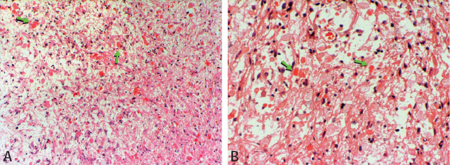
Discussion
A
pilocytic astrocytoma is the most common pediatric central nervous system
glial neoplasm, and the most common pediatric cerebellar neoplasm. Pilocytic
astrocytomas usually present within the fi rst two decades of life. The
cerebellum, optic nerve, optic chiasm, and hypothalamic regions are the
favored locations for the tumor, however it can also be found within
the cerebral hemispheres, ventricles, and spinal cord. When the lesion
is reported in a cerebral hemisphere, the temporal lobe is the most common
site. The nature and duration of the patient’s symptoms is related to
the specific location of the tumor category. |
|
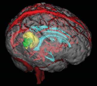
Figure
2 (above)
Neuronavigation image: nodule in
green and cyst in yellow
|
Classical imaging features include a mixed cystic and solid mass with
avid enhancement of the solid component of the mass. This appearance
is seen in two-thirds of cases. A mass with enhancement of the cyst wall
in conjunction with the mural nodule, necrosis with non-enhancing portion
of the mass, and a predominantly solid mass without a cystlike component,
are the less common imaging features of this neoplasm. Peri-tumoral edema
is rarely noted. Surgical resection is the treatment of choice for this
neoplasm with up to 79 % twenty-year survival. Radiation and chemotherapy
is reserved for recurrent tumors, and tumors situated within the optic
chiasm and hypothalamus.
References Koeller
KK
& Rushing EJ. From the Archives of the AFIP: Pilocytic Astrocytoma:
Radiologic-Pathologic Correlation. RadioGraphics 2004; 24: 1693-1708 |