ASTROCYTOMA
DRS.
F. DISCEPOLA,
M.C. GUIOT,
L. SOUALMI
AND A.
OLIVIER
|
A |
29-year-old
right-handed woman with no past medical history who developed
nonspecific neck pain and headaches subsequent to a low speed
motor vehicle accident. |
|
Her
symptoms led her to consult the emergency room at a remote hospital.
At the time of her presentation, she denied experiencing seizures. Her
physical exam demonstrated no neurological deficits. All blood work
obtained was unremarkable. She underwent a nonenhanced CT scan, which
demonstrated a mixed solid and cystic mass lesion residing within the
right parietal lobe. The patient was then referred to a neurosurgeon
at the Montreal Neurological Institute for further imaging and
assessment. |
 Imaging
Findings Imaging
Findings
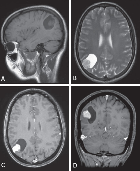
Magnetic resonance imaging characterized the lesion as a single 3.5
x 3.7 x 2.6 cm intraparenchymal mixed solid and cystic mass lesion located
wi thin the right parietal lobe. There was no peri-tumoral associated
vasogenic edema or necrosis, nor was there ring enhancement of the cystic
portion of the mass. The solid component followed iso T1 and T2 signal,
and enhanced
avidly post gadolinium infusion. (Figure 1)
Figure 1
(A) Sagittal T1 image without gadolinium. (B) Axial T2 image.
(C) Axial T1 post gadolinium. (D) Coronal T1 post
gadolinium. Both gadolinium images show avid enhancement of the solid
portion of the mass lesion. |
|