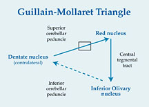
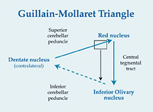
If the lesion is located in a cerebellar hemisphere and involves the dentate nucleus, the olivary degeneration will be contralateral, due to the decussation of the dentato-rubral fibers.
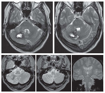
(<) Fig 5 MR images obtained in a patient with caveroma at right dentate nucleus tat bled Axial T2-weighted image shows
|
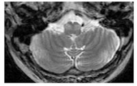
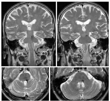
Fig 6 (>) If the lesion involves the superior cerebellar peduncle, the degeneration will occur contralaterally and a paramidline lesion, if located near the peduncle, may result in bilateral HOD, if there is involvement of both the dentato-rubral fi bers, and the CTT.
|
|
 REFERENCES REFERENCES1) Uchino A., Hasuo K., Uchida K. et al. (1993) Olivary degeneration after cerebellar or brain stem hemorrhage: MRI. Neuroradiology 35: 335-338 2) Kitajima M., Korogi Y., Shimomura O., et al. (1994) Hypertrophic olivary degeneration : MR imaging and pathologic findings. Radiology 192: 539-543 3) Rieder C., Rebouças R., Ferreira M. (2003) Holmes tremor in association with bilateral hypertrophic olivary degeneration and palatal tremor. Chronological considerations. Arq Neuropsiquiatr 61: 473-477 4) Goyal M., Versnick E., Tuite P, et al. (2000) Hypertrophic olivary degeneration: Metaanalysis of the temporal evolution of MR fi ndings. AJNR 21: 1073-1077 5) Birbamer G., Buchberger W., Felber S., et al (1992)MR appearance of hypertrophic olivary degeneration: temporal relationships. AJNR 13: 1501-1503 6) Goto N., Kaneko M. (1981) Olivary enlargement: chronological and morphometric analyses Acta Neuropathol (Berl) 54: 275-282 |
|
8