| Hypertrophic
olivary degeneration (HOD): an unusual way to degenerate Drs. Fabrizio Venturi, Donatella Tampieri, Roland Brassard & Denis Melanson |
|
|
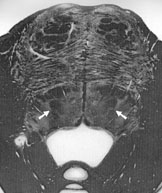
In the CNS the degeneration of an anatomical structure is usually characterized by neuronal loss replaced by proliferation of glial elements. This change is reflected in the appearance on MRI: a loss of volume associated with a hypersignal on long TR sequences. There is, however, a case in which the degeneration is accompanied by hypertrophy: the degeneration of the inferior olivary nucleus, described for the first time in 1887 by Oppenheim from anatomic specimens. Not seen on CT scan due to artefacts caused by bony structures, the hypertrophic olivary degeneration (HOD) is more recently detected in vivo by MRI and is characterized by an hypersignal of the olive of the medulla oblongata on PD/T2 images with a variable enlargement of the olive itself. |
|
|
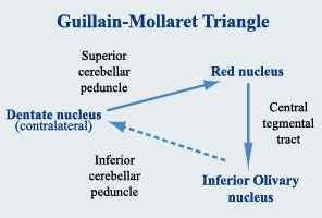
HOD is considered a trans-synaptic degeneration because it occurs following loss of neuronal input to a cell, in this case the neurons of the inferior olivary nucleus. HOD occurs when a lesion, usually a haemorrhage, causes an interruption of the Guillain-Mollaret triangle (Fig1). |
 Fig1 |
|
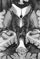
The Guillain-Mollaret triangle is a triangular circuit connecting the dentate nucleus of the cerebellum of one side with the red nucleus and the inferior olivary nucleus of the other side, via the superior cerebellar peduncle and the central tegmental tract (CTT) (Figs. 2, 3). |
|
 |
Fig 2
|
 |