|
"A small hurt
in the eye is a great one" – Old English proverb
Dedicated
imaging of the orbits can provide a wealth of information on a
wide variety of lesions seen in the eye and adjacent structures.
CT, with or without intravenous contrast, can be used to image
bone and soft tissues and is especially useful in the setting of
trauma. Axial images are obtained with a slice thickness of 2 mm
with coronal and sagittal reformats.
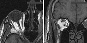
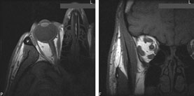
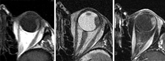
MRI is especially useful in cases
of orbital mass lesions. It is routinely performed using a head
coil with images acquired in the axial, sagittal and coronal
planes, with T1, T2 and post-gadolinium fat saturated sequences.
A study is currently underway at the MGH comparing the
conventional head coil to the more sensitive surface coil, which
increases spatial resolution.
|