|
A Fourth ventricle meningioma
Drs. Mohammad AbuRemsh, Marie-Christine Guiot & Denis Sirhan |
|
|
The patient is a 66 year-old lady who had a 4 year history of headache associated with nausea and vomiting, more recently having gait difficulty and balance problem. She was diagnosed and treated at the onset as Meniere's syndrome. The headache was generalized, not following certain time nor activity. Eventually a CT scan was done which showed a tumour in the region of the 4th ventricle and she was referred for MRI and surgery. On neurological examination, she was ataxic with a tendency to fall to the left, but she had no cranial nerve dysfunction, more specifically no swallowing difficulty nor dysphonation. MRI displayed a well delineated tumour within the lower 4th ventricle which could be totally removed. Histology of the lesion was typical for meningioma. |
|
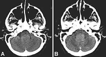 |
CT plain (A) and enhanced (B) showing smooth round lesion in
4th ventricle |
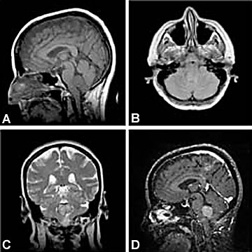
A) Sagittal T1; lesion slightly hypointense to adjacent brain
(B) Axial Proton Density; slightly hyperintense to adjacent brain
(C) Coronal T2; minimal more hyperintensity than on PD with rim of hypointensity
(D) Sagittal Gradient T1; lesion enhances moderately with Gado. |
 |