| Images of "Moya-Moya" arteritis
Roland Brassard M. D. And Daniel Gendron, M. D. |
|
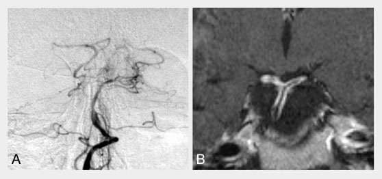
A 37 year old male who developed in early August 2000 severe headaches with mild right hand numbness and clumsiness.When seen in E.R. he had a mild right hemiparesis with right hand sensory loss and a right homonymous hemianopsia.. Initial imaging showed an infarct in left posterior cerebral territory and other areas of ischemia in left frontal lobe. A lumbar puncture was done and normal. He was initially treated with high dose IV steroid on the assumption of an aggressive cerebral vasculitis. Despite steroids, a few weeks later, he developed more severe hemiparesis and he underwent a non-dominant temporal lobe meningeal biopsy which was unhelpful, not showing vasculitis. A trial of IV steroid with monthly IV cyclophosphamide was attempted, but despite this, his state worsened and he got completely hemiplegic on the right and aphasic. He has been, in the spring of 2001, treated with a bilateral cerebral vascular bypass procedure to improve cerebral blood flow. We have considered his disease to be a form of "Moya-Moya"-type arteritis. (A) frontal view of arterial phase of right vertebral angiogram: note the narrowing of the lumen of the basilar artery, and more severely of the right posterior cerebral artery. Compare with the appearance of same vessels on (B) coronal T1-weighted image with Gadolinium: the residual lumen is flow/void while the arterial wall is enhancing. |