|
AN
INNOVATIVE PHOTOGRAPHER
Charlie
showed great interest and staying power for developing new photographic
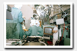
techniques. In the 1950s, he helped Dr. Cone with the use of the
Knebel 35-mm strobe-light camera, which gave the surgeons convenient
play by play records of their operations.
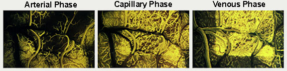
But his best known innovation was the
invention in the mid-1960s of the highly specialised photographic
technique for recording in the Cone laboratory and in the operating
room the blood flow in the surface vessels of the brain. This
procedure, which we called fluorescein angiography of the brain
(FAB), could only have been created with Charlie's energy and
persistence in grappling with formidable technical problems.
|
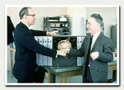
William Feindel and Charles
Hodge greet each other in front of a model of the radioisotope
method for measuring blood flow during brain surgery
(1969).
|
|
Although
the fluorescence of the dye flushing through the brain looked
brilliant, the amount of light to expose the film was actually
quite low. With ingenious originality, he processed Kodak 35mm
colour film with ASA of 160, the fastest at that time, to obtain
the equivalent of 1500 and eventually up to 4000. He also cleverly
worked out in immense detail the use of complex colour filters.
|