by standard
MR imaging in as many as 20% of subjects. The addition
of diffusion weighted MR sequences to our protocol
increased the number of positives to 33% (5/15).
Future questions we wish to pursue
are the following; Do these imaging findings predict clinical
course? Will the signal changes resolve with resolution
of symptoms? What is the exact nature of the lesions detected
on delayed DWI but not spin-echo T2? Do these findings
correlate with other investigative findings such as baseline
and full neuropsychology battery? Is there a correlation
with neuropsychological and neurophysiological testing?
(Dupuis et al. Neuroreport, 2000, Leclerc 2001 in prep
). Do imaging findings reflect repeated or cumulative
injury? Our future prospective studies will address these
important issues in our attempt to improve the diagnosis
and management of sport related head injury.
Address correspondence
to
Karen M. Johnston, MD, Ph.D; Director of Neurotrauma,
Department of Neurosurgery; Montreal General Hospital,
1650 Cedar Ave., Montreal, Quebec, CANADA H3G 1A4
(514) 934-8062
|
|
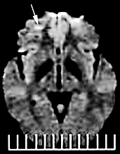
Diffusion-MR
image showing a small subcortical right frontal hyperintense
focus. Note that the lesion is distinct from frontal artefact
seen adjacent to bone. |
|
 Axial-T2 image of the brain showing abnormal hyperintense
foci in the sub-cortical white matter of the frontal and
posterior parietal lobes.
Axial-T2 image of the brain showing abnormal hyperintense
foci in the sub-cortical white matter of the frontal and
posterior parietal lobes.