|
Transcranial Doppler (TCD) is a simple and readily available means of exploring the arteries at the
base of the cranium and provides hemodynamic information that is complementary to other diagnostic
imaging method such as MRA and CTA. The classical indications include the detection and follow-up of
vasospasm in subarachnoid hemorrage, detection of intracranial stenosis, and assessment of intracranial
collaterals. Monitoring intracranial hypertension, brain death, cerebral emboli are other potential indications
currently under clinical evaluation.
The recent development of ultrasound contrast agents has improved significantly Doppler
studies and opened the door to new applications by adding anatomical information to hemodynamic data.
These contrast agents contain air microbubbles sufficiently small to cross the pulmonary capillaries and to
recirculate in the systemic circulation, allowing good intravascular enhancement for both imaging and spectral
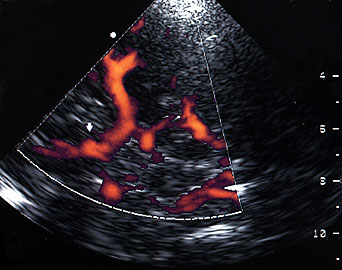
analysis. We can now study intracranial aneurysms, as small as 3 mm in diameter (fig1). Coupling contrast
agents with 3D power Doppler is a very promising technique for this indication. With this technique, it is
also possible to study arteriovenous malformations, such that we can evaluate the feeding arteries, nidus
and draining veins.
Given its high prevalence, investigation of arteriosclerotic disease, by assessing intracranial
stenosis, collateralization or distal hemodynamic consequences of a stenosis, will still remain the first indication
of TCD. The use of contrast agent permits a good evaluation in the majority of patients, and TCD is a
normal complement to cervical Doppler in this clinical setting.
|